12 Lead EKG
The 12 lead EKG is a widely accepted medical standard for electrocardiogram administration in healthcare facilities across the United States and around the World. While the term ‘lead’ is often used loosely to refer to the cables that are attached to the body through which signals are transmitted to the monitoring device, it actually references the tracings that result due to voltage differentials. This can become a bit confusing considering that it is common to use ten cables to conduct a 12 lead EKG. The lead placement has been standardized in the US.
Electrode Number and Placement
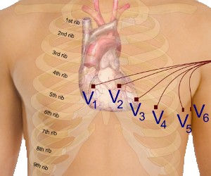
The standard electrocardiogram uses ten cables that are connected to different parts of the patient’s body via a gel that conducts electricity and an adhesive pad. There is an electrode for the right arm (RA), left arm (LA), right leg (RL), left leg (LL), 2 for the fourth intercostal space on either side of the sternum (V1 and V2), 1 for the fifth intercostal space at the midclavicular line (V4), one that goes between V2 and V4 (V3), one for the left anterior axillary line (V5), and one for the midaxillary line (V6). Welch Allyn has created an informative visual representation of proper lead placement that can be found by clicking here. As they point out, for optimal results it is important to make sure the patient is warm and relaxed, the areas for electrode placement have been shaved and cleaned, and the skin has been given time to dry before the electrode is attached.
12 Lead EKG Waves and Intervals
The tracings that are created by the administration of the electrocardiogram consist of waves and intervals that are used to evaluate the electrical conduction in the heart and determine if abnormalities exist. There are many different types of heart diseases that can cause deviations of the baseline tracing and the identification of each depends on the technician’s ability to understand and appreciate a normal tracing. Technician training programs and certification exams are meant to help individuals when learning about the fundamental waves and intervals characteristic of a standard electrocardiogram so that they can provide an initial review of the exam before sending it off to be evaluated by the members of the advanced medical team.
The three most important waves seen on a 12 lead EKG are the P wave, QRS complex, and T wave. There is another wave (U wave) that is sometimes seen, but is often concealed by the T wave. To understand the significance of each of these waves it is important to appreciate that electrical conduction in the heart begins at the top of the atria and travels down through the ventricles signaling cardiac contraction as it moves from one point to the next. Electrical signals are created within an area of the heart that is known as the sinoatrial node. This area is a small collection of tissue that is located near the top of the right atria. A similar collection of tissue located at the convergence of the four heart chambers is referred to as the atrioventricular node. The P wave represents the atrial depolarization that occurs as the electrical signal travels from the sinoatrial node toward the atrioventricular node.
 The QRS complex is a dramatic spike from baseline that is seen on the standard 12 lead EKG as the electrical signal travels from the atrioventricular node through the right and left ventricles. The size of the QRS complex is reflective of the fact that the ventricles contain far more mass than the atria. The T wave follows after a QRS complex on the electrocardiogram and it is representative of the return of the cells within the ventricles to a resting state. Some professionals in the industry refer to this stage as the refractory period.
Intervals are segments between separate points on waves that allow the healthcare professional to evaluate conductive attributes that may indicate that a heart condition exists. The most important intervals seen on the standard 12 lead EKG include the RR interval, PR interval, ST interval, and QT interval. The RR interval is the segment between two sequential R waves and it can be used to evaluate the resting heart rate. A normal rate is between 60 and 100 beats per minuted. The PR interval is the segment between the beginning of a P wave and the beginning of the sequential QRS complex. This interval is used to evaluate the conduction of electrical impulses from the sinoatrial node through the atrioventricular node. The ST interval is a segment that is measured from the J point to the end of the T wave. The QT interval is a segment that begins at the start of the QRS complex and ends at the end of the sequential T wave. The QT interval is often used to assess patient’s who are suspected of having ventricular tachyarrythmias that could lead to sudden cardiac death.
Relevance For EKG Technicians
Administration of the standard 12 lead EKG is a common responsibility that a technician has. A technician is expected to become proficient in lead placement and the basic evaluation of the tracing quality. Employers have developed training programs that address these topics in detail and test the technician’s understanding of the topics prior to the provision of patient care. In addition, many administrators require new employees to pass a nationally recognized certification examination that can demonstrate competence in the industry.