What is an Echocardiogram
The echocardiogram is a procedure that uses sound waves to create images of the heart. To collect these images, the technician places an instrument called a transducer on the chest of the patient. The transducer emits waves that penetrate the body and get reflected back. As the transducer receives the reflected waves, it transmits them to a computer where they are converted into moving images. Physicians use the echocardiogram images with other studies to evaluate the heart and determine whether or not any abnormalities exist.
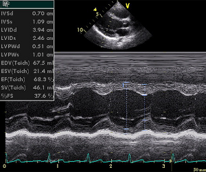
Transthoracic Echocardiogram
There are a few different types of echocardiograms that can be used to evaluate the heart. A transthoracic echocardiogram is a common approach because it is easy to perform and usually does not cause discomfort. To perform a transthoracic echocardiogram, a technician places the transducer on the chest and adjusts it so a high quality image can be captured. Cardiologists often use this type of echo to evaluate abnormal heart sounds such as clicks and murmurs, create an image of an enlarged heart, assess the cause of pains or shortness of breath, and determine what is causing irregular beats. In addition, a transthoracic echo may be performed for many other reasons including the need to measure the thickness of the heart wall, evaluate the valves, measure the size of the chambers, calculate the amount of blood flowing through the heart, and much more.
Stress Echocardiogram
 The stress echocardiogram is a little more complex than a transthoracic echo because it collects images before and after the heart has been stressed. This approach can cause discomfort for patients because they have to exercise or have a medication injected that increases the heart rate. The stress echo evaluates coronary artery disease through the detection of a decrease in blood flow to the heart. Cardiologists order a stress echo when they think that a patient has suffered myocardial ischemia (decrease in the level of oxygen being delivered to the heart cells).
Doppler Echocardiogram
The Doppler echocardiogram can be performed while any of the other three types of echocardiograms are being administered. Doppler technology is used to evaluate how blood circulates through the heart chambers, valves, and blood vessels. It can show the speed and direction of blood flow as well as add color to the image to help evaluate important parameters.
Transesophageal Echocardiogram
The transesophageal echocardiogram requires that the probe be inserted into the esophagus rather than be placed on the chest wall. While this type of echocardiogram requires a patient to be sedated, it provides images of the heart that are not obstructed by the lungs and ribs. The transesophageal echo can be used during surgery to monitor heart function and is great for evaluating if valves are functioning as they should. This procedure is also used to find clots in the heart, evaluate flow between chambers, and guide cardiac catheterizations.